Friday, November 4, 2011

2.75 Urine


recall that urine contains water, urea and salts.
-brain influences the function of the 2 kidneys.
-the 2 kidneys are filters our blood.
-the molecules which are removed which come down the ureter collects in the bladder and together these form urine.
-urine contains salts, water, urea.
-salt and water affects the composition of tissue fluid which is called osmoregulation.
-the removal of urea is part of the process of the excretion of metabolic waste.
-composition of urine (salts, water and urea) varies and depends on the condition which a person is operating.


2.74 ADH.


describe the role of ADH in regulating the water content of the blood.
-ADH- anti-diuretic hormone.
-ADH is produced in the region of the brain called the hypothalamus, it flows through the bloodstream and the target is the kidney.
-the effect of ADH is to control and alter the composition or the quantity of water in the blood.
-ADH can make the blood more or less concentrated- tissue fluid is isotonic to the cytoplasm of the cell.


-ADH targets the collecting duct- allows more water to come out of the collecting duct.
-collecting duct- where water is selectively reabsorbed into the blood.
-it is possible to increase the amount of water going into the blood by applying ADH.
-ADH makes the collecting duct balls have more pores- more water can escape from the collecting duct which goes back into the blood.
-the consequence of ADH secretion is that the urine coming from the collecting duct will be more concentrated and it will have a lower volume. (because of all the water escaping from the ADH)


2.73 Glucose reabsorption


understand that selective reabsorption of glucose occurs at the proximal convoluted tubule.
-selective- means that the molecule is selected (glucose)
-reabsorption- go from the glomerular filtrate and it will go back into the blood. remove the molecule from the blood and then put it back.
-filtration occurs in the bowman's capsule.
-items in the filtrate- glucose, water, salts and urea.
-water is removed back into the blood in the collecting duct.
-the end of the nephron is the urine and normally urine does not contain glucose.
-if you test urine for glucose and receive a positive test- indication of condition called diabetes.
-but yet there is glucose in the fluid at the start- in the bowman's capsule- this is because in the proximal convoluted tubule (proximal- first, convoluted- twisted, tubule- tube) section glucose is removed and is taken back into the blood.
-glucose is selectively reabsorbed into the blood in the proximal convoluted tubule.

2.72 Water re-absorption.


understand that water is reabsorbed into the blood from the collecting duct.
-the bowman's capsule- location of ultra filtration.
-blood comes into the kidney placed under high pressure and the dissolved content of the blood are forced into the bowman's capsule tube- glomerular filtrate (contains glucose, water, salts and urea)
-as the filtrate passes a long the tubular and when it reaches the collecting duct (vertical tube) water is removed from the filtrate. the water will go back into the bloodstream. water has been selected and reabsorbed into the blood- called selective reabsorption.
-selected reabsorption of water occurs in the collecting duct.

2.71 Ultrafiltration


describe ultrafiltration in the Bowman's capsule and the composition of the glomerular filtrate.
-nephron of the kidney- structure which carries out the filtration of the blood.
-results in filtered blood (clean blood) with waste products removed (urine)
-urine composed of water (the majority) salts and urea (molecule which contains nitrogen waste from body- toxic)
-urine comes out into the pelvic region and drains into the bladder.
-urine is produced at the beginning of the nephron called bowman's capsule (filtration process begins) known as ultra filtration.


-the filtration of blood begins with the blood arriving in the kidney in the nephron in the blood vessel called the afferent arteriole- the blood is coming in (high pressure blood from artery) (wide blood vessel)
-the twisted and coiled bit is called the glomerulus.
-the blood vessel that comes out of the bowman's capsule the diameter is smaller. (narrow blood vessel)
-the blood pressure increases in the glomerulus. the consequences is that the high pressure forces the liquid within the blood (plasma-component which dissolves in blood eg. water as solvent, salts, glucose, amino acids, urea.)- these things are all forced out of the blood vessel and into the space- inside of the bowman's capsule. 
-when the plasma is forced inside the bowman's capsule we change the name to filtrate. and because it is in the glamourous we call it the glomerula filtrate instead- contains water, salts, amino acids, glucose and urea.
-blood has been filtered by pressure due to the smaller restricted diameter of the blood vessel, generating a high pressure which forces the liquid to the tube.


2.70 Nephron Structure


describe the structure of the nephron to include the Bowman's capsule and the glomerulus converted tubules, Loop of Henle and the collecting duct.


-nephron- functional unit of the kidney. part that does the filtration and the controlling of the composition of the blood.
-branch of aorta- taking blood into the kidney is the renal artery.
-kidney filters the blood and the contents which are removed from the blood (excreted) are called urine which comes down the structure called the ureter and collects in the bladder for release.
-the filtered blood exits in the renal vein and returns to the vena cava.

-if we slice the kidney in half to show the internal structure we see different colour regions.
-lighter colour is called the cortex.
-darker colour is called the medulla.
-the space in the middle (lighter colour) is called the pelvic region. space is where the urine collects and drains down the ureter.

-the reason for the different colours is because the kidneys is made up of many million tubilar structures tubes.
-the tubes starts on the edge of the medulla and moves directly upwards through the medulla and out into the cortex and dips into the medulla again and back up and comes to a dead end.
-the dead end is called the bowman's capsule.
-the structure is called the nephron.


-above the dotted line is the cortex and below the dotted line is the medulla and the dotted line where the tube ends is the pelvic (where urine emerges).
-the tube is made up of twisted sections called the convoluted tubules.
-duct= tube.
-the dip is called the loop of Henle. (dips into the medulla)
-the dead end cup shape structure is called the bowman's capsule.
-the tight knot of blood vessels in the bowman's capsule is called the glomerulus.
-the first twisted section is called the proximal convoluted tubules (PCT)
-the second twisted section is called the distal convoluted tubules (DCT)
-the arrangement of this nephron structure which gives the different colour regions in the kidney.
-there are millions of nephrons in a single kidney.


Monday, October 31, 2011

2.69 Urinary system


describe the structure of the urinary system, including the kidneys, ureters, bladder and the urethra.
-2 kidneys- right and left kidney- each with its own blood supply carry out the process of excretion and filtration and osmoregulation.
-from each kidney there is a tube that leads to the bladder- called the ureter- carries the urine from the kidney to the bladder.
-there is a common bladder for both ureters- urine is conducted from the outside of the body through the urethra down through the vagina or through the penis.


2.68b Osmoregulation


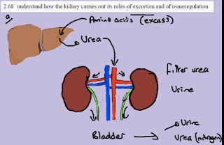
understand how the kidney carries out its role of excretion and of osmoregulation.
-osmo: osmosis
-regulation: control

-the fluid which surrounds the cell- must be isotonic with the cytoplasm of the cells- the water going in and out of is equal and the cells will remain the same size and the same shape and maintain their function.

-the danger to the tissue is that blood circulating into the tissue would be concentrated causing a hypertonic tissue fluid (remove to much water) or it may be very dilute causing a hypotonic tissue fluid (add too much water).

-so we want to keep the tissue fluid isotonic to the cells cytoplasm- achieved by controlling the composition of blood.

-blood forms the tissue fluid- role of kidney to control the composition of blood.

-blood with circulates through our kidney excess water, salts can be removed and excreted down the ureter.

-by controlling the content of water and salts in the blood the kidney can keep the blood and therefore the tissue fluid isotonic with the cells cytoplasm maintaining the function of the cell.

2.68a Excretion


understand how the kidney carries out its role of excretion and of osmoregulation
-excretion of the molecule urea (carries nitrogen and it is toxic to the body and cannot be stored)
-the original form of nitrogen circulating in our blood stream and is potential toxic are the amino acids.
-amino acids are normally used for growth but extra amino acids must be removed- they are toxic, rule of both liver and kidney.
-first stage- blood circulates to the liver and the amino acids are broken down and converted into the molecule urea.
-the urea re-enters the bloodstream and circulates to both kidneys.
-the kidneys will filter the urea from the blood and the urea will be added to the water to form urine.
-urine drains down the ureter to collect in the bladder.
-the urea is now in the bladder in the form of urine- been removed from the body.
-the filtered blood returns to the circulation in the veins with the toxic amino acids and urea removed- example of excretion.

Sunday, October 30, 2011

2.67b Human organs of Excretion


recall that the lungs, kidneys and skin are the organs of excretion.
-1) lungs- lungs excrete carbon dioxide- waste from respiration that need to be eliminated from the body.
-2) kidney- kidneys excrete excess water, the molecule urea (the nitrogen waste from amino acids- we cannot store them so we excrete them) and salts.
-3) skin- known to excrete water (sweat), salts (sweat), and a little bit of urea. (mainly water and salt)

2.67a Excretion in plants


recall the origin of carbon dioxide and oxygen as waste products of metabolism and their loss from the stomata of a leaf
-photosynthesis: absorbing light energy and in the process of doing so it combines (CO2) carbon dioxide and water (H2O) to form glucose (C6H12O6) and gives of the gas (O2) oxygen- waste molecule example of excretion which is the release of metabolic waste.
-process of respiration: glucose (C6H12O6) + O2 (oxygen) --enzyme reaction-----> ATP (produced in the process) + CO2 (waste molecules also excretion) + H2O.

-plants excrete O2 (oxygen) or CO2 (carbon dioxide) depending on whether they are doing photosynthesis or respiration.

Wednesday, October 12, 2011

3.31 Evolution


describe the process of evolution by means of natural selection.
-evolution: change in the form of organisms. (new form of organisms arising)
                : change in frequency (how many) of alleles.
-natural selection: the mechanism of evolution (first proposed by Charles Darwin)
-SA (Staphlococcus aureus) causes skin infection and also lung infection


evolving SA

first graph
-normal form is susceptible to methicillin (antibiotic) (antibiotic can kill SA)
-MSSA (methicillin susceptible SA)

second graph
-random mutation in SA allow breaking down of methicillin. 
-because it can break down it can no longer killed by the antibiotic (resistant form)
-MSRA (red on graph)

-2 forms of bacteria (called evolution) [MSSA, MSRA]
-when antibiotic are applied to the population MSSA is decreased and MRSA is becoming increasing common
-evolution: increase in frequency of allele (or resistance) 


2 features
-random mutation produces MRSA form
-the non random selection (due to antibiotics) selects MRSA to survive and MSSA to be selected and killed.
-natural selection is a process not a list.


Tuesday, October 11, 2011

3.34 Causes of mutation


understand that the incidence of mutations can be increased by exposure to ionizing radiation (for example gamma rays, X-rays and ultraviolet rays) and some chemical mutagents (for example tobacco)
-radiation (ionizing radiation) eg. x-rays and UVB (ultraviolet) rays (sunshine) can cause skin cancer.
-due to chemicals (tars in tobacco) cause mutation to base sequence producing new alleles and causes cancer.
-chemicals that cause mutations are called mutagens.
-mutagens which also causes cancer are called carcinogens.

3.33 Antibiotic resistance


understand how resistance to antibiotics can increase in bacterial populations.
-bacterial population is Staphlococcus aureus- known to cause skin infection and lung infection.
-if infected can be treated with drug called methicillin (type of antibiotic)
-type of chemical will kill the SA (Staphlococcus aureus)
-the type of SA which is killed by the methicillin is called the susceptible form (can be abbreviated to MSSA) (MSSA- methicillin susceptible Staphlococcus aureus)

-there was a random mutation to SA. when the methicillin (antibiotic) was applied the bacteria did not die (resistant form)
-the type of SA which is resistant to the methicillin is abbrivated to MRSA (MRSA- methicillin resistant Staphlococcus aureus)
-the mutation has create genes in SA, when expressed, allows it to break down the antibiotic and resists which causes bacteria to not die.
-if antibiotic is used across time the form of bacteria increasing survives, becomes more common and this becomes a serious problem in hospitals because antibiotic no longer work.

3.32 Types of mutation


understand that many mutations are harmful but some a neutral and a few are beneficial.


- gene ---mutation---> new alleles (responsible for phenotypes)
-new alleles can be beneficial (eg. improve efficiency of enzyme), harmful (eg. non functional enzyme), or no effect (neutral effect)- may not last forever and with the environmental change the mutation can become harmful or beneficial.

3.30 Mutation


recall that mutation is a rare, random change in genetic material that can be inherited.


-form of gene is called the allele.
-certain processes can cause a change in the base sequence which creates a new version of the allele. (possible that the allele will result in the production of an entirely different protein and have an entirely different effect on the phenotype)
-different alleles exist because of mutation. (Allele A and Allele a)
-mutation changes base sequence of the gene.

3.29 Species Variation


understand that variation within in a species can be genetic, environmental or a combination of both.

-variation- difference in phenotype of individuals. it is possible to count or measure and shown in graphic form.
-individual has a phenotype, appearance of an individual is because of their genotype, modified by the environment.
-variation in population is the variation of the individuals.
- Vpopulation= Vgenotype + Venvironment    (V- variation)

1. -different classes or group, this suggests variation in the population (which is variation in the spices) is entirely due to the variation in the genotype. environment is playing no role.
-eg. blood groups (A, AB, B, O)


2.
- variation in population, variation in the spices- causes by genetic variation (variation in genotype) but the different group is modified to form the smooth curve (distribution curve) caused by the environment.
-eg. height (quality of your diet [environment])
-continuous variation


3. 
-variation in population or species (phenotypic variation) entirely due to environmental variation. genes have no role.
-eg. home language
-cannot be inherited.

Tuesday, September 20, 2011

3.10 Menstrual Cycle


understand the roles of oestrogen and progesterone in the menstrual cycle.
-oestrogen and progesterone are examples of hormones.
-hormones are produced in a structure called and endocrine gland.
-hormone will travel through the blood from the endocrine gland to target tissue.
-at the target tissue the hormone will have an effect. (may have multiple effects)
-ovary is the endocrine gland for oestrogen, it produces oestrogen which flows in the bloodstream to the lining of the uterus where it has effect no.1 which is the thickening of the endometrium. effect no.2 is that it flows to the bloodstream to the brain and it releases a second hormone known as luteinising hormone (LH) the production of luteinising hormone reaches its peak at around day 13- causes the ovary to release an egg into the oviducts where it is possible for fertilisation.

-in ovary- during the first half of the menstrual cycle the follicle becomes larger and larger- inside follicle is an egg. the cells around the follicle is producing oestrogen.
-the follicle reaches it largest point at around day 13 when the luteinising hormone causes the wall of the outside to rupture and the egg is released. - called ovulation.
-once process is complete the empty follicle changes function and colour (to yellow) gives us the name corpus lutetium (known as the yellow body) this yellow body is known to produce progesterone.
-ovary and the corpus lutetium in particular is the endocrine gland for progesterone.

-progesterone travels through the blood stream to the lining of the uterus to give effect no.3 to maintain the lining of the uterus- prevents the lining of the uterus from breaking down.
-in this condition it is possible that a fertilized egg could implant to the wall of the uterus and develop into a pregnancy.
-but if there is no implantation of an fertilized egg the corpus lutetium breaks down and progesterone levels fall.

-the fall in progesterone levels lead to effect no. 4
-which is the breakdown in the lining of the uterus- menstrual period/menstrual bleeding.
-marks end of one menstrual cycle and a start of a new menstrual cycle.
-when the lining has broken down completely the cycle and the change in hormones begins one more.


Saturday, September 17, 2011

2.4 Plant and Animal Cells


describe the differences between animal and plant cells.

ANIMAL
-has a nucleus, cytoplasm, and has a cell membrane like a plant cell.
-but it stores glycogen (a type of carbohydrate) as the molecule and it has an irregular shape.

PLANT
-has a nucleus, cytoplasm, and has a cell membrane like an animal cell
-but it has a cell wall, chloroplasts, large central vacuole and is in a regular shape.
-plant cell stores the molecule starch (a type of carbohydrate).